
EG-UR5 Echoendoscope
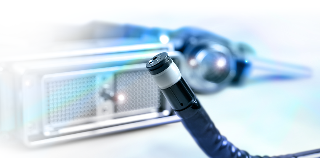
The SonoScape EG-UR5 brings endoscopic visualization and 360° radial ultrasound together in a single advanced platform for evaluating the upper gastrointestinal tract and surrounding anatomy. Designed to look beyond the mucosal surface, the EG-UR5 combines conventional endoscopic imaging with ring-array ultrasound scanning across a broad 4.5–12.5 MHz frequency range, giving physicians the ability to examine the GI wall, submucosal structures, and adjacent organs from within the gastrointestinal tract. Its clinical applications include TN staging of upper digestive tract malignancies, evaluation of submucosal lesions, and assessment of pancreatic, biliary, gastric, duodenal and other surrounding pathology. With a 140° endoscopic field of view, 360° ultrasound scanning, a 2.2 mm instrument channel, four-way angulation and balloon or water-based acoustic coupling, the EG-UR5 provides a versatile diagnostic EUS solution designed to integrate SonoScape endoscopy and ultrasound into one clinical workflow.
Highlights
- 360° Radial Ultrasound Imaging: Ring-array ultrasound technology provides a complete 360° view around the distal tip, supporting detailed evaluation of the GI wall, submucosal structures, and surrounding anatomy. The EG-UR5 operates across a broad 4.5–12.5 MHz frequency range for flexibility across different imaging depths and clinical applications.
- Endoscopy and Ultrasound in One Scope: The EG-UR5 combines a 140° endoscopic field of view with radial EUS, allowing physicians to transition from conventional upper-GI visualization to ultrasound assessment of structures beyond the mucosal surface during the same examination.
- Designed for Diagnostic EUS: Developed for applications including TN staging of upper digestive tract malignancies, evaluation of submucosal lesions, and assessment of pancreatic and biliary disease, the EG-UR5 extends visualization beyond what can be seen with conventional gastroscopy alone.
- Versatile Imaging and Procedural Access: The EG-UR5 supports B, M, CFM, PW, PDI and THI ultrasound modes, along with a 2.2 mm instrument channel, four-way distal angulation, and balloon or sterile-water acoustic coupling—providing a flexible platform for comprehensive diagnostic EUS examinations.
| Specification | EG-UR5 |
|---|---|
| Distal Diameter | 11.5mm |
| Channel Diameter | 2.2mm |
| Working Length | 1250mm |
| Angulation | Up: 180° | Down: 90° | L/R: 100° |
| Field of View | 140° |
| Depth of View | 3-100 |
| Ultrasound Frequency | 4.5-12.5 MHz |
| Ultrasound Modes | B, M, CFM, PW,PDI, THI |
See The Image Quality For Yourself
- Broadband EUS Imaging: The EG-UR5 operates across a 4.5–12.5 MHz frequency range, providing flexibility to balance penetration and image detail when evaluating the GI wall and surrounding anatomy.
- Complete 360° Visualization: Ring-array scanning produces a full 360° radial ultrasound view, providing a circumferential perspective of the GI wall, submucosal structures, and adjacent anatomy.
- Advanced Ultrasound Imaging Modes: The EG-UR5 supports B, M, CFM, PW, PDI, and THI imaging modes, providing multiple approaches to tissue and vascular assessment during EUS examinations.
- Optimized Near-Field Imaging: Balloon or sterile deaerated-water acoustic coupling provides direct acoustic contact at the target anatomy, supporting detailed ultrasound visualization from within the upper GI tract.
GET THE 411 – Up to the Second -> Straight To Your Inbox